Fluoroscopy And C Arm Is Utilised In A Wide Range Of Diagnostic And Therapeutic Treatments9/29/2022 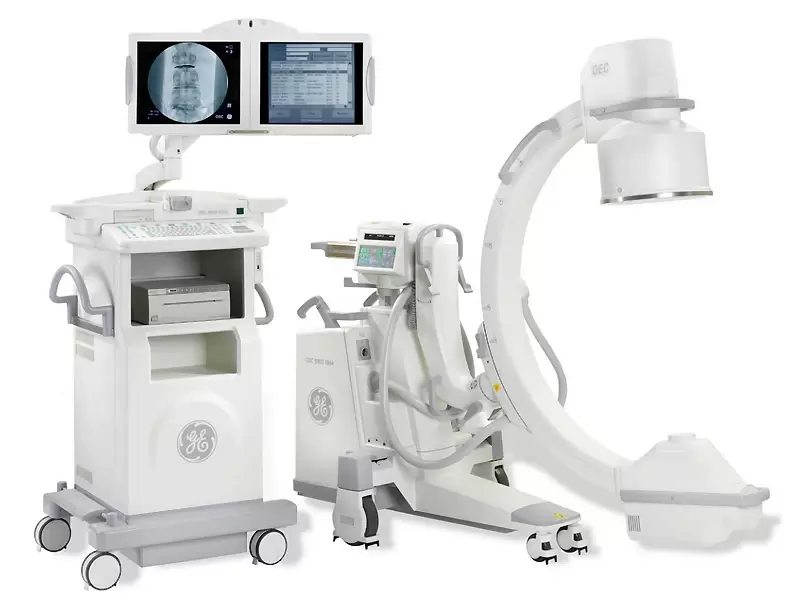 Fluoroscopy and C-arm Market With the aid of the imaging technique known as fluoroscopy, doctors may view internal organs, muscles, and bones in real-time images of the body. This allows them to make important surgical decisions while causing the least amount of discomfort to the patient. Fluoroscopy And C Arm initially employed image intensifiers, but modern versions use flat panel detectors for digital image processing, which greatly lowers the radiation dose given to the patient.X-ray technology is the basis for the operation of C-arm imaging systems. X-ray source and detector are the two main parts of C-Arms. High resolution real-time X-ray images are provided by C-Arms, which aid surgeons in monitoring the procedure, correcting problems as they arise, and minimising the need for additional surgeries.
According to its association with an electronic equipment that amplifies light and converts it into video signalling to present an electronic display, fluoroscopy is described as an imaging method utilised for imaging and producing visible electronic images of patients for therapy.Medical imaging techniques like Fluoroscopy And C Arm is utilised in a wide range of diagnostic and therapeutic treatments. Fluoroscopy offers X-ray imaging, which is particularly helpful for examining the interior makeup and bodily operations of patients. The most frequent applications for it are in interventional radiology, general radiology, and image-guided surgery. The C-arm technique is also based on X-ray technology and is often utilised in orthopaedics, cardiology, traumatology, and vascular surgery. The C-shaped arm that joins the X-ray source and X-ray detector is where the term "C-arm" originates. Fluoroscopy And C Arm is an imaging method that employs X-Ray technology to capture live images of the body, enabling medical professionals to see inside organs, muscles, and bones. Fluoroscopy equipment processes digital images using flat panel detectors to minimise the radiation dose given to the patient. A fluoroscopy and C-arm is a tool that doctors use to direct a needle to a particular spot with a live X-ray screen. The image detector and X-ray generator on a mobile C-arm are the two components of a fluoroscopy system. Similar to X-ray imaging, Fluoroscopy And C Arm is employed in the study of human body parts to provide real-time images. These tools are essential for orthopaedic surgery, cardiac catheterization, lumbar puncture, biopsies, inserting interventional catheters into arteries and veins, and interventional radiology treatments.The C-Arm is a portable imaging device that is primarily used for fluoroscopic imaging during orthopaedic and surgical operations. The two main parts of a C-arm are an X-ray source and an X-ray detector. C-arms operate on the principles of X-ray technology. High resolution real time X-ray images from C-Arms enable surgeons monitor surgery, make significant changes when necessary, and lessen the likelihood of needing to perform additional surgery. Doctors can undertake minimally invasive surgical treatments because to fluoroscopy.
0 Comments
Leave a Reply. |
