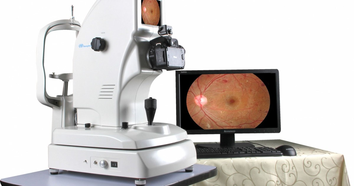
 Fundus Camera A fundus camera is a device that captures images of the retina. These images can be used to diagnose ocular conditions and document alterations in the eye. Optometrists, ophthalmologists, and trained medical professionals use Fundus Camera to perform examinations and screening programs. They also use these devices in research studies to assess changes in the retina and to report observations.
The glaucoma diagnosis is an important part of glaucoma management, as it allows the detection and prevention of glaucoma. The diagnosis can be made in community centers, hospitals, or optometry clinics. Fundus imaging is a common ophthalmic imaging modality that can be easily performed in many settings, including communities and remote sites, such as villages and isolated communities. The low cost of the camera enables the use of portable Fundus Camera that can be used in less-developed countries to perform population-based screening for glaucoma. Several AI-based approaches have been developed to detect glaucoma, and these techniques generally involve the automated processing of segmented fundus images. They may either use binary classification or multi-class classification. Eye doctors uses a range of tools to diagnose age-related macular degeneration. These include the visual acuity test, dilated eye exam and Amsler grid. The doctor also might use a test that shows tiny yellow deposits called drusen under the retina. This is an early sign of the disease and can help a doctor determine if they have macular degeneration. Another useful tool is fluorescein angiography. This uses a dye that moves through the blood vessels in the retina and takes pictures as it passes through. Fundus photography is a diagnostic tool that can help identify cataracts. This eye disease occurs when the crystalline lens loses its transparency and impedes the ability to see clearly. This condition can lead to severe visual loss and blindness, especially in older adults. Therefore, the early diagnosis of cataracts is essential to improve patients’ quality of life. Fundus Camera imaging can be used to evaluate and document abnormalities related to disease processes affecting the eye, as well as to follow progress of certain conditions such as macular degeneration, retinal neoplasms and choroid disturbances. It may also be used to evaluate the effect of recent surgery or to assess the therapeutic response of a treatment, such as surgery for glaucoma. Fundus photography is also known as retinal photography, is the process of taking pictures of the eye using a special camera. The photographs are used to assess the health of the optic nerve, vitreous, retina and blood vessels. Fundus photography is a valuable tool in the diagnosis of retinal vascular diseases like diabetic retinopathy, glaucoma, macular degeneration and other conditions. It can help the doctor identify the stage of the disease at that moment in time and track its progression. Fundus Camera technology has made it possible to perform ocular imaging on patients without an ophthalmologist, which is especially important in the emergency department and other fast-paced settings where time is of the essence. Nonmydriatic tabletop and portable fundus cameras, as well as smartphone-based devices, offer non-physician staff a practical and cost-effective means of screening for neurologic disorders with ocular abnormalities
0 Comments
Leave a Reply. |
