 Intraoperative Imaging is an optical imaging technology often used by surgeons to get timely and in-depth image of surgical site during the surgery. The technique provides surgeons image of organ, cell, or tissue, on which surgeon perform surgery. Mobile C-arm, intraoperative X-ray, intraoperative MRI, intraoperative CT, and intraoperative ultrasound are some commonly used intraoperative imaging devices or techniques; used in different surgical procedures such as cardiac surgery, neurosurgery, cancer surgery, and gastrointestinal surgery. Intraoperative technologies are designed to produce better patient outcomes and reduce iatrogenic injury, and thus, the use of these technologies has increased worldwide.
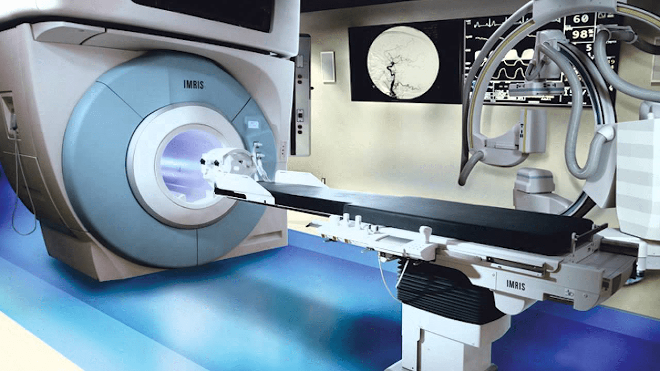
An intraoperative CT scanner brings this technology into the operating room, allowing doctors to sync existing scans with new ones. Having access to all of this information at once allows surgeons to better make critical decisions during delicate surgeries, such as those involving the brain. Intraoperative Imaging modality that provides interactive and timely information during surgical procedures. Because the transducer is in direct contact with the organ being examined, high-resolution images can be obtained that are not degraded by air, bone, or overlying soft tissues. While, intraoperative magnetic resonance imaging (iMRI) is a procedure that creates images of the brain during surgery. Intraoperative Imaging is often used in conjunction with surgical navigation systems, similar to a GPS device. These techniques help surgeons determine exactly where to perform surgery, how much tissue to remove, and where to make cuts. The technology also allows surgeons to visualize changes in the brain as surgery proceeds. It also helps detect misplaced screws and areas of dangerous bleeding. Moreover, intraoperative imaging is becoming a standard tool in cancer surgery. Advances in technology are rapidly advancing. Some of the advancements will result in a significant change in the operative approach and introduce new surgical techniques. This in turn will help improve patient outcomes. There is no limit to how Intraoperative Imaging will improve the practice, and in the meantime, there are already many promising applications in the field. Surgeons rely on iMRI technology to obtain accurate pictures of the brain that guide them in removing brain tumors and treating other conditions such as epilepsy. MRI is especially helpful for imaging the brain. To utilize MRI technology during surgery, surgeons use special imaging systems and operating rooms, including: Portable iMRI devices, which are moved into the operating room to create images. Magnetic resonance imaging (MRI) is also used to investigate or diagnose conditions that affect soft tissue such as tumors or brain disorders. Intraoperative imaging is a rapidly expanding field encompassing many applications that use a multitude of technologies. Intraoperative Imaging offer faster results, simplified consoles, high image quality, and easier operations as compared to traditional imaging devices. In terms of imaging, one of the major differences between diagnostic and intraoperative imaging is in the fact that during intervention the surgeon only needs the relevant physiological and anatomical information for dynamic monitoring of the surgical process. Imaging is one of the pillars for the ongoing evolution of surgical oncology toward a precision paradigm.
0 Comments
Leave a Reply. |
