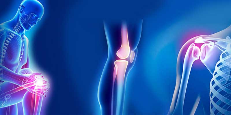
 Orthopedic Medical Imaging Orthopedic Medical Imaging refers to the usage of X-rays, Magnetic Resonance Imaging, and Computed tomography to examine diseases and wounds that impact the bones, muscles, tendons, ligaments, cartilage, and spine. Professional Orthopedists can further utilize these test outputs to make an appropriate treatment strategy for their patients. X-Rays are most usual kind of imaging utilized in orthopedics is the X-ray, which sheens a small quantity of radiation from the organ of the body being diagnosed.
The Orthopedic Medical Imaging is known to increase in several parts across the globe because of the rising and developing usage of developed medical infrastructure and increasing necessity of medical processes. Rising graph of the geriatric population is also considered to be a reason for the growth of orthopedic medical imaging. Orthopedic Medical Imaging offers diagnostic analysis for a range of MS issues. These tests consist X-ray, MRI, and CT scans. X-rays are usually utilized to measure bone fractures, soft cell wounds, and joint fitness. They are a rapid, suitable, and painless process that enables physicians to observe the severity of damage that has occurred in the body. X-rays are best at showing the 2 D of a specific body part and can aid physicians see more bone detail compared to that which Magnetic Resonance Imaging can provide. Often, an X-ray is the only method to analyze a disease or wound. Musculoskeletal Ultrasound is other exceptional analyzing tool, musculoskeletal ultrasound is conducted with an ultrasound device that creates pictures of muscles and joints on the body. This can identify even the slight cut in an individual’s muscles and ligaments that a Magnetic Resonance Imaging might not show. Bone Scan is an X-ray of the joint that is conducted to observe and cure specific concerns such as cracks and inflammation. The scan can show if a bone has recovered totally and can be supportive in curing the injured part. Magnetic Resonance Imaging utilizes very potential magnets to generate high-resolution pictures of the bones and soft cells. It also includes a computer to gather slices of the Magnetic Resonance Imaging pictures into a descriptive 2D pictures of an affected organ of the body. Computed Tomography scans are alike to X-rays however they offer a very descriptive picture of the body. Anyhow, they can be time consuming to attain the results and may expose individuals to more radioactivity compared to X-rays. Magnetic Resonance Imaging (MRI) is a very particular diagnostic devices compared to X-rays and Computed Tomography scans as it takes 3-D images of the joints and other cells in the body. It also do not expose individuals to the hazardous radiation of X-rays or Computed Tomography scans. Magnetic Resonance Imaging can be utilized to plan procedure on injured bones, particularly those that have ruptures or deformities. This decreases time in the OT and enhances interaction amidst the medical staff. Orthopedic physicians are instantly developing ML designs to forecast and cure orthopedic diseases based on a patient’s MRI and other test outputs. These models have a high diagnostic perfection compared to physical examination only, which makes them appreciated tools for individuals and orthopedic professionals similar. Fluoroscopy is an extra kind of diagnostic imagery test that aids physician see the internal body parts. It can be utilized to detect difficulties such as a cardia arrest, IBD or a cancer. In orthopedics, Magnetic Resonance Imaging and Computed
0 Comments
Leave a Reply. |
